
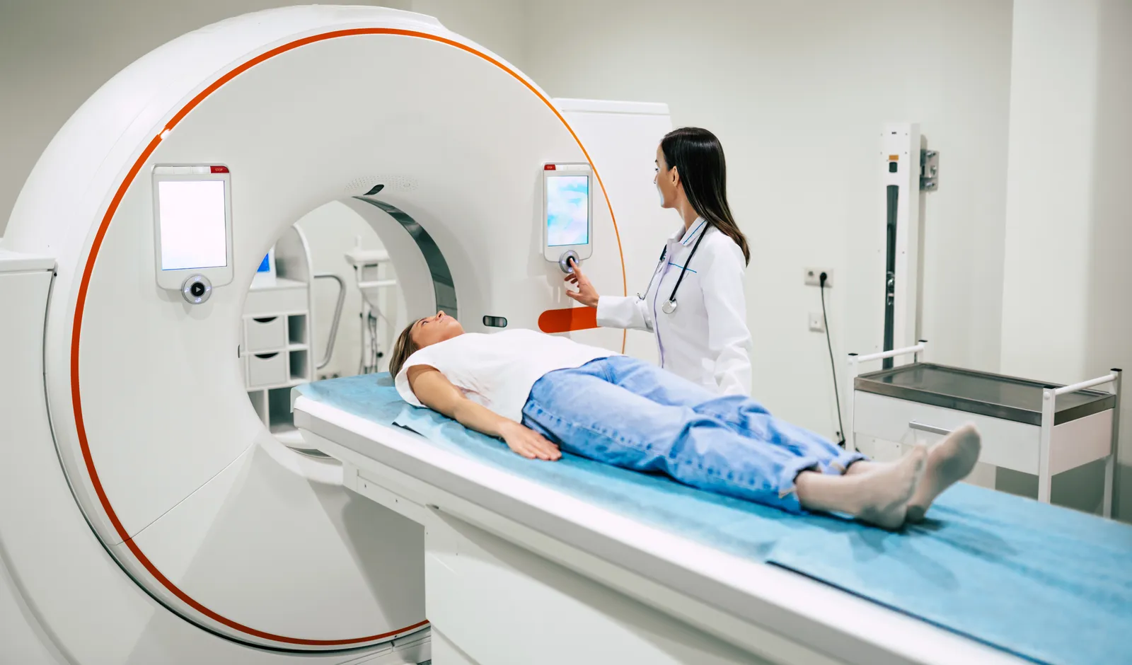
A coronary CT angiogram (CCTA) is a non-invasive scan that gives you a detailed picture of the arteries that supply blood to your heart. It can show plaque in the artery walls, where the arteries have narrowed, and how severe the narrowing is.
This test is also called coronary CT angiography, coronary CTA, CT coronary angiography, cardiac CTA, or CTCA. It is non-invasive, meaning no catheter travels through an artery to your heart. However, it requires an IV and iodine-based contrast and uses a small to moderate dose of radiation.
Currently, the American Heart Association guidelines recommend CCTA for evaluating certain people with chest pain or suspected coronary artery disease. It is not the right first test for every person, and it is not a replacement for lowering cholesterol, high blood pressure, smoking, diabetes, or other established risks.
What is a coronary CT angiogram (CCTA)?
A CT coronary angiogram (CTCA) uses a fast CT scanner to take many X-ray images of your beating heart. A computer combines those images into detailed views of the coronary arteries. These are the blood vessels that carry oxygen-rich blood to the heart muscle.
In this test, contrast is injected through an IV in your hand or arm. The contrast makes blood inside your arteries appear bright, which helps a radiologist see the artery walls and signs of narrowing. RadiologyInfo describes CCTA as a CT examination of the coronary arteries using iodine-containing contrast.
CCTA and a catheter angiogram are different procedures. During a catheter angiogram, a doctor threads a tube through an artery in the wrist or groin to the heart. A catheter angiogram can allow a doctor to evaluate blockages and place a stent during the same procedure. CCTA takes 3D pictures of the heart; it does not treat blockages.
What does a CCTA show?
CCTA can show whether you have visible plaque, where it sits, and how much it narrows the channel for blood. It can also show the course and shape of your coronary arteries.
What CCTA can show
| Finding | What it means |
|---|---|
| No visible plaque | The scan did not find plaque large enough to see in the coronary arteries. |
| Noncalcified plaque | Plaque without enough calcium to appear on a calcium score scan. It is sometimes called soft plaque. |
| Calcified plaque | Plaque that contains calcium. It can make an artery look brighter on CT. |
| Stenosis | The percentage of narrowing in the channel where blood flows. |
| Plaque burden | The overall amount and spread of plaque, which is different from the main narrowing. |
| Plaque location | The same plaque can matter more in the left main artery than in the right artery. |
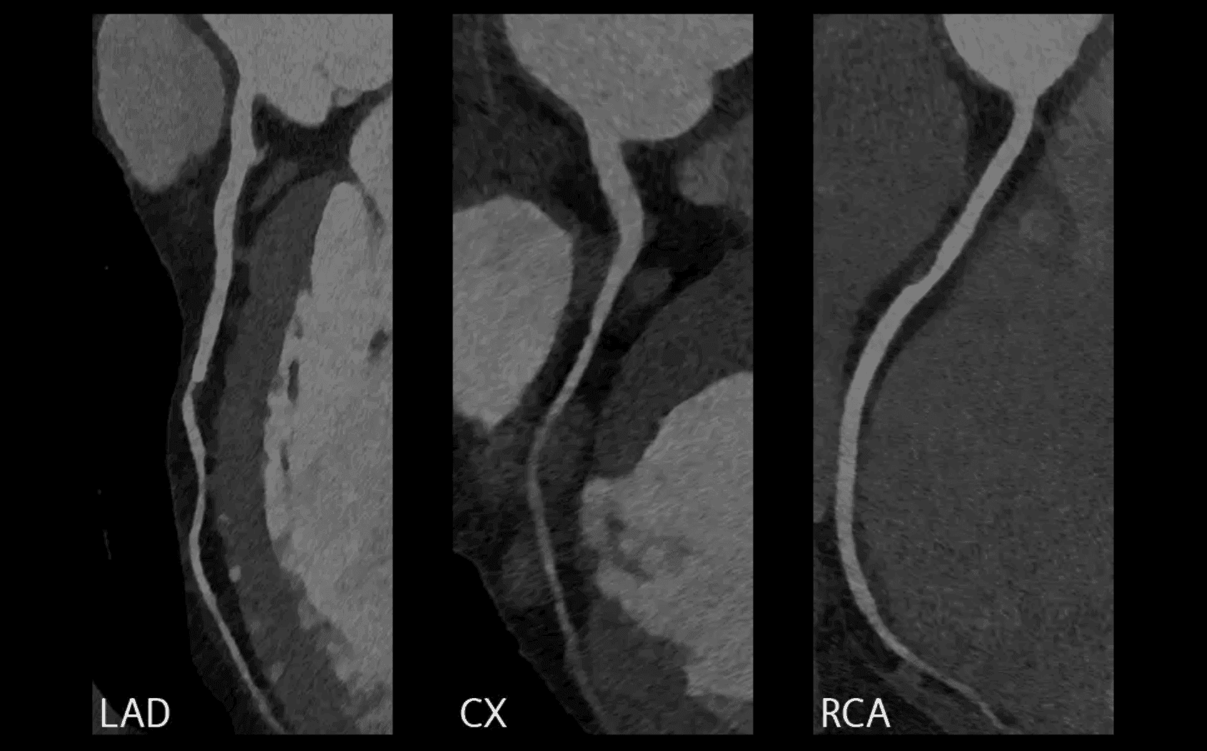
Calcified, noncalcified, and mixed plaque
Plaque is not simply risky soft plaque and harmless hard plaque. It may be calcified, noncalcified (soft), or mixed. It may also show features linked with higher risk:
- Low-attenuation plaque: An area of high-risk, low-density plaque.
- Spotty calcification: Small calcium deposits within plaque.
- Positive remodeling: Outward expansion of the artery wall around plaque.
- Napkin-ring sign: A ring-like pattern within plaque.
These features can add information about risk, but they have to be considered with the total plaque burden, narrowing, symptoms, and the rest of the report. The CAD-RADS 2.0 consensus uses the term high-risk plaque because no scan can identify the exact plaque that will cause a future heart attack.
Plaque burden is not the same as blockage
A person can have a large amount of plaque spread across several arteries without one severe blockage. Another person can have less total plaque but one tight narrowing. Both matter: about two-thirds of heart attacks start in plaque that narrows the artery by less than half. This is why a useful CCTA report describes both plaque and stenosis.
Plaque begins in the artery wall. As it grows, the wall can expand outward and temporarily preserve the channel for blood. Doctors call this positive remodeling. A CCTA can often see plaque and outward remodeling even when there is little narrowing, although very small or subvisual plaque may remain below the scan's resolution. The 2025 American College of Cardiology scientific statement defines visible coronary plaque as abnormal tissue in the artery wall with or without extension into the channel.
What can CCTA not tell you?
A CCTA gives you detailed images, but it cannot capture every process inside an artery. A normal scan means the radiologist found no plaque large enough to see at the scan's resolution. It cannot rule out microscopic plaque.
- It cannot tell you whether a specific plaque will rupture.
- It cannot always tell whether a moderate narrowing reduces blood flow enough to cause symptoms.
- It does not measure all causes of chest pain, such as small-vessel disease, inflammation around the heart, or many lung and digestive problems.
- It does not show the heart's electrical rhythm the way an ECG does.
- It cannot replace your symptoms, medical history, blood pressure, cholesterol tests, or a clinician's judgment.
When a narrowing is uncertain, a doctor may use a computer analysis of blood flow called fractional flow reserve from CT (FFR-CT), a CT blood-flow test called CT perfusion, or a stress test. When symptoms continue despite no major blockage, another evaluation may be needed for small-vessel disease or a noncoronary cause.
Who may need CCTA?
CCTA works best when it answers a clear clinical question. For example, it can help your doctor determine whether coronary artery disease may be causing your chest discomfort when you do not already have known obstructive disease.

- Stable chest pain with an intermediate or higher likelihood of coronary disease. A CCTA can look for plaque and narrowing when symptoms are ongoing but not an emergency.
- Intermediate-risk acute chest pain after an initial evaluation. In an emergency department, CCTA may be one option after an ECG of the heart's electrical activity, troponin blood tests for heart muscle injury, and a structured risk assessment.
- An unclear or inconclusive stress-test result. CCTA may help show whether an artery has an anatomical narrowing.
- Known nonobstructive coronary disease with continued symptoms. A clinician may use CCTA or another test when the result could change care.
- A suspected coronary artery abnormality. CCTA can map where the arteries begin and the path they follow.
The 2021 American Heart Association chest pain guideline says CCTA is useful for diagnosing and estimating risk in people with stable chest pain who have an intermediate to high likelihood of coronary disease and no known disease. It also says low-risk people may not need urgent testing.
Can you get a CCTA without symptoms?
Currently, guidelines do not recommend CCTA as a routine screening test for every adult without symptoms. Doctors more often use a calcium score to decide whether you should start a preventive medication like statins.
There are selected cases where a clinician may consider CCTA because noncalcified plaque is a particular concern, including some higher-risk younger adults. The 2026 American Heart Association guideline discusses selective use rather than broad screening. A decision should consider whether the result is likely to change prevention, along with the contrast, radiation, cost, and possibility of follow-up testing.
Imaging should not become a reason to delay care for an established risk factor. For example, a person with very high LDL cholesterol may already qualify for treatment based on guidelines and personal history. A scan can show a picture of the heart arteries, but it does not erase years of exposure to plaque-causing cholesterol particles.
When is CCTA generally not recommended?
Doctors often choose a different test in the following situations because CCTA may be unsafe, produce unclear images, or delay care you need urgently.
- Possible heart attack symptoms. Seek emergency care instead of scheduling an outpatient CCTA. An emergency team can check your ECG and blood tests immediately and move to a catheter angiogram if you need urgent treatment.
- Severe kidney disease or a recent kidney injury. Your kidneys filter iodine contrast from your blood. When they have less reserve, contrast exposure may add to the risk of further injury. Your doctor may check a blood test called eGFR and weigh that risk against the benefit of the scan.
- A previous reaction to iodine-based contrast. A prior reaction raises your chance of another one. Your clinician and imaging center may give you medicine beforehand, change the contrast plan, or choose another test. The American College of Radiology contrast manual explains how clinicians make this decision.
- Pregnancy or possible pregnancy. CCTA exposes a fetus to radiation. Your clinician and imaging center will first decide whether you need the scan now and whether another test can answer the question without radiation.
- A fast or irregular heart rhythm, or trouble holding your breath. The scanner must freeze a moving heart. An uneven heartbeat or body movement can blur the arteries, although medicine and newer scanners can help in some cases.
- A large amount of coronary calcium. Calcium can create a bright blur that hides the channel inside an artery or makes it look narrower than it is. Newer photon-counting scanners and calcium-artifact reduction may help, but they are not available everywhere.
How is CCTA different from other heart tests?
The question each heart test answers
| Test | Main question | Key difference |
|---|---|---|
| Calcium score | How much calcified plaque is present? | No IV contrast. It does not show soft plaque or directly measure narrowing. |
| CCTA | Is visible plaque present, and how narrow are the arteries? | Uses IV contrast and can show calcified, noncalcified, and mixed plaque. |
| Stress test | Does exercise or medicine reveal reduced blood flow or another stress-related problem? | It tests heart function, not plaque buildup in arteries. |
| Catheter angiogram | What does the channel inside the arteries look like, and is treatment needed now? | It is invasive. Doctors mainly use it to check significant narrowing and place a stent when needed. |
| ECG | What is the heart's electrical activity at that moment? | It shows the heart's electrical activity, not plaque buildup in arteries. It can help detect an ongoing heart attack. |

No one test is the most complete test for every question. A calcium score may be enough for a prevention decision. A stress test may be better when the main concern is blood flow during exertion. A catheter angiogram may be appropriate when severe disease is already likely and treatment may be needed. A whole-body MRI, such as a Prenuvo scan, doesn't evaluate the coronary arteries, so it can't replace these tests.
Read moreCCTA vs calcium score: which heart scan should you get?How do you prepare for CCTA?
Your imaging center may give you different instructions based on its scanner and your health. Follow its instructions. A typical checklist includes:

- Keep your heart rate steady. Avoid regular and decaf coffee, tea, other caffeine, and nicotine for 24 hours. Avoid exercise before the scan on the day of your appointment.
- Take heart-rate medicine only as prescribed. Heart-rate-lowering medicine can bring your heart rate into the desired range, often near or below 60 beats per minute. This can reduce the time you spend at the imaging center and improve image quality.
- Avoid medicines that can interact with nitroglycerin. If you are taking Viagra, stop 48 hours before. If you are taking Cialis or Levitra, stop 78 hours before. Confirm the timing with your imaging center, and do not stop a prescription medicine without instructions from the clinician who manages it.
- Prepare for contrast. Tell the center about kidney disease and previous contrast reactions. If you are older than 60 or have kidney risk factors, complete any kidney-function blood test the center orders before your appointment.
- Prepare for the appointment. Do not eat for two hours before the scan, drink two eight-ounce glasses of water during that time, wear clothes without metal, and arrive 30 minutes early. Allow roughly 90 minutes for the visit.
These steps help staff at your imaging center capture clear pictures safely. A slower, steadier heartbeat reduces motion. Hydration supports normal kidney clearance of contrast. A short fast can reduce nausea, and avoiding certain medicines prevents a dangerous interaction with nitroglycerin.
What happens during CCTA?
- A technologist places sticky ECG patches on your chest to track your heartbeat.
- An IV is placed in your hand or arm for the contrast.
- Staff at the imaging center may give you a beta blocker and nitroglycerin if they are appropriate for you.
- You lie on a table that moves through a short, open CT scanner.
- The technologist asks you to stay still and hold your breath for a few seconds.
- The contrast injection may cause a brief warm feeling or a metallic taste.
- The scanner takes the pictures while your ECG helps time them to your heartbeat.
The scanner can collect the images in seconds. Your full appointment may take an hour or longer if staff at the imaging center need time to prepare you or lower your heart rate. Mayo Clinic notes that heart-rate medicine can add time to the visit.
Most people can return to usual activities after a scan. The facility may ask you to wait briefly, especially if you received medicine. Follow its instructions about driving, hydration, exercise, and when to restart any medicine that was held.
Is CCTA safe?
Most people complete CCTA without a serious problem. Before the scan, staff at the imaging center check the factors that affect safety and image quality, including your kidney function, previous contrast reactions, blood pressure, heart rate, pregnancy status, and current medicines.
Contrast risks
Staff at the imaging center use iodine contrast to make the blood inside your arteries stand out. Most people tolerate it well. Tell your doctor if you previously reacted to CT contrast because that history helps the imaging center plan the scan. A shellfish allergy by itself does not mean you are allergic to iodine-based contrast.
Your kidneys filter iodine contrast from your blood. Severe kidney disease or a recent kidney injury leaves them with less reserve, so contrast exposure may add to the risk of further injury. Your doctor will review your eGFR, which estimates kidney function, along with your hydration, medicines, other health conditions, and how urgently you need the scan.
Radiation
CCTA uses ionizing radiation, but the protocol often matters more than the scanner's model name. Prospective ECG triggering turns on the X-rays during only a selected part of your heartbeat. Studies report about 60 to 80 percent less radiation than older retrospective gating, which keeps the X-rays on through more of the cardiac cycle.
How CCTA technique can change radiation dose
| Technique | Reported dose or reduction |
|---|---|
| Older retrospective ECG gating | About 8 to 20 mSv in older machines |
| Prospective ECG triggering | About 60 to 80 percent lower than retrospective gating |
| Lower tube voltage with prospective triggering | Reviews report reductions ranging from 3 to 83 percent, depending on voltage and protocol |
| Photon-counting CT | Can reach below 1 mSv in selected research protocols |
These figures come from different studies and patient groups. You cannot add the reductions together or use the table to predict your personal dose.
A systematic review of lower-voltage prospective CCTA found a wide range of doses and reductions because body size, voltage, tube current, and reconstruction method all affect the result. Ask your imaging center for the estimated radiation dose for your scan.
Why you may receive medicine and an IV
A beta blocker slows your heart so the scanner can capture sharper pictures. Nitroglycerin briefly widens your coronary arteries so the radiologist can see them more clearly. Because both medicines can lower blood pressure, staff at the imaging center check your blood pressure, health history, and current medicines before giving them.
How accurate is CCTA?
CCTA is especially useful for ruling out major obstructive coronary disease in appropriately selected patients. A high-quality scan with no visible plaque or narrowing is strongly reassuring.
How do you read a CCTA report?
Start with the impression at the end of your report. It should summarize whether the scan found plaque, the worst narrowing, which arteries it affects, and whether the radiologist could not evaluate any part of the scan.
Many US cardiac imaging programs use CAD-RADS 2.0, the current version of the Coronary Artery Disease Reporting and Data System. The main category runs from 0 for no visible plaque or narrowing to 5 for at least one completely blocked artery. CAD-RADS 2.0 also adds an overall plaque-burden category and letter modifiers.
CAD-RADS 2.0 in plain English
| Category | General meaning |
|---|---|
| CAD-RADS 0 | No visible coronary plaque or narrowing. |
| CAD-RADS 1 | Minimal narrowing, from 1% to 24%. |
| CAD-RADS 2 | Mild narrowing, from 25% to 49%. |
| CAD-RADS 3 | Moderate narrowing, from 50% to 69%. |
| CAD-RADS 4A | Severe narrowing of 70% to 99% in one or two coronary arteries. |
| CAD-RADS 4B | At least 50% narrowing in the left main artery or at least 70% narrowing in all three major coronary arteries. |
| CAD-RADS 5 | At least one completely blocked coronary artery. |
| CAD-RADS N | The scan or an important segment could not be fully evaluated. |
The letter P followed by a number describes overall plaque burden from P1, mild, through P4, extensive. Other modifiers may note high-risk plaque features, stents, bypass grafts, or blood-flow analysis. The CAD-RADS 2.0 document says you should read the category with the full report rather than use it by itself to choose treatment.
Use a free toolOpen your CCTA files in Veevo's DICOM viewerWhat happens after the results?
Your next step depends on why you had the scan and what it found. Review the report with a clinician who can consider your symptoms, family history, blood pressure, cholesterol, blood sugar, smoking history, and current treatment.

No visible plaque
A normal CCTA is reassuring and can make major coronary narrowing unlikely. It does not mean future risk is zero or that prevention no longer matters. Blood pressure, cholesterol exposure, smoking, diabetes, exercise, sleep, and family history still affect long-term risk.
Nonobstructive plaque
Nonobstructive coronary disease means plaque is present but no artery has a major blockage. It is still coronary atherosclerosis. A clinician may discuss lowering cholesterol, controlling blood pressure and blood sugar, avoiding tobacco, exercise, food choices, and whether medicine is appropriate. Do not wait for a plaque to become a severe blockage before taking established risks seriously.
Moderate or severe narrowing
If your report shows CAD-RADS 3 or 4A, your doctor may use FFR-CT, including HeartFlow, CT perfusion, or a stress test to learn whether a narrowing reduces blood flow.
CAD-RADS 4B generally leads to a catheter angiogram because it means at least 50 percent narrowing in the left main artery or severe disease in all three major coronary arteries. CAD-RADS 5 means a total blockage. It may be new or old and may require urgent care.
Read moreHeartFlow analysis: what it is, cost, and who needs itAI plaque analysis
Some companies offer AI analysis of your CCTA images. Cleerly is one example. The AI can measure how much plaque you have, where it sits, and how much appears calcified or noncalcified. This information may help your doctor assess risk when the standard CCTA already shows visible plaque, but not every scan includes AI analysis, and researchers have not yet shown that adding it improves outcomes. The 2025 American College of Cardiology statement says a physician should review both the original images and the AI output.
Read moreCleerly heart scan: what it is, cost, and is it worth it?How much does CCTA cost?
The price varies by location, facility, insurance, and what the service includes. Hospitals often post gross charges around $2,000 to $3,000, and some published hospital prices exceed $5,000. These are sticker prices, not necessarily what your insurer pays or what you owe.
Your insurer may cover CCTA when a clinician orders it for a supported reason, such as evaluating certain chest-pain symptoms. Your plan negotiates its own rate, then applies your deductible, copay, or coinsurance. Preventive CCTA without symptoms may be less likely to qualify for coverage, and your plan may require prior authorization.
Before you schedule, ask for a written estimate that includes the scan, contrast, heart-rate medicine, radiologist's interpretation, and any optional FFR-CT or AI plaque analysis. Confirm that both the facility and interpreting doctor participate in your insurance network.
The Veevo Heart Scan is for $1,199, including referral and scheduling, the scan, a board-certified radiologist read, and a doctor results review.
Veevo Heart ScanClinician-reviewed. HSA/FSA eligible. Learn more.How do you get a CCTA?
You usually need an order from a licensed clinician. The clinician should explain what question the scan will answer for you and confirm that you can safely receive contrast and heart-rate medicines. Some services arrange the clinical review, imaging order, scheduling, and results consultation for you.
Choose a center that regularly performs cardiac CT. Ask about its scanner, dose-reduction methods, heart-rate preparation, and who interprets the scan. Also ask how the center will send you the report and original image files, often called DICOM files.
The bottom line
CCTA can show you visible plaque in the walls of your heart arteries, the overall spread of disease, and how much the arteries have narrowed.
The scan has its limits. It uses contrast and radiation, image quality varies, and a narrowing does not always tell you whether blood flow is reduced. You will get the most value when you and your clinician have a clear reason to use CCTA, an experienced center performs it, and your doctor interprets it with the rest of your health information.
14 Sources
American College of Cardiology 2021 AHA/ACC chest pain guideline perspectives
https://www.acc.org/Latest-in-Cardiology/ten-points-to-remember/2021/10/27/14/06/2021-Guideline-for-Chest-Pain-gl_chestpainAmerican College of Cardiology Quantitative coronary plaque analysis in clinical practice: 2025 ACC scientific statement
https://www.jacc.org/doi/10.1016/j.jcmg.2025.11.008American College of Radiology ACR Manual on Contrast Media
https://www.acr.org/Clinical-Resources/Clinical-Tools-and-Reference/Contrast-ManualAraki et al. Ultra-low-dose coronary CT angiography using photon-counting detector CT
https://pmc.ncbi.nlm.nih.gov/articles/PMC11631184/Blumenthal et al. 2026 guideline on the management of dyslipidemia
https://www.jacc.org/doi/10.1016/j.jacc.2025.11.016Cardia Vision Patient coronary CT angiogram instructions
https://sa1s3.patientpop.com/assets/docs/321308.pdfCury et al. CAD-RADS 2.0: 2022 coronary artery disease reporting and data system
https://pubs.rsna.org/doi/10.1148/ryct.220183Falk et al. Coronary plaque disruption
https://pubmed.ncbi.nlm.nih.gov/7634481/Hospital Ledger CPT 75574 hospital cash and gross prices
https://www.hospitalledger.com/procedure/75574Mayo Clinic CT coronary angiogram: about, preparation, and results
https://www.mayoclinic.org/tests-procedures/ct-coronary-angiogram/about/pac-20385117Radiological Society of North America and American College of Radiology Coronary CTA
https://www.radiologyinfo.org/en/info/angiocoroctShen et al. Prospective ECG-triggered coronary CT angiography: clinical value of noise-based tube current reduction with iterative reconstruction
https://pmc.ncbi.nlm.nih.gov/articles/PMC3669389/Siemens Healthineers Evaluate even heavily calcified vessels with photon-counting CT
https://www.siemens-healthineers.com/en-us/computed-tomography/ct-technologies-and-innovations/pure-lumenTan et al. Low tube voltage prospectively ECG-triggered coronary CT angiography: a systematic review
https://pmc.ncbi.nlm.nih.gov/articles/PMC6209486/