Written by Miles Witte
Published
Trace 400 years of heart disease detection, from blood circulation and the first ECG to cardiac catheterization, echocardiography, MRI, and CT.
Written by Miles Witte
Published
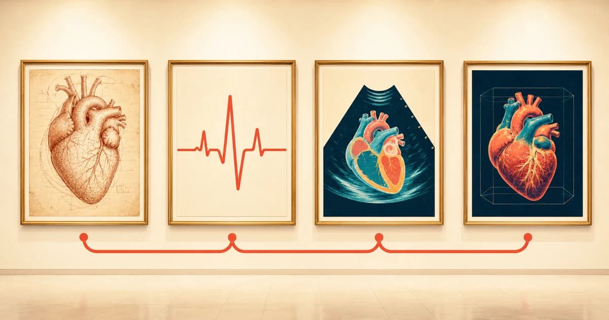
Heart disease detection has changed dramatically over the past 400 years. Physicians moved from studying how blood travels through the body to recording the heart's electrical signals, measuring pressures inside it, and creating detailed images of its muscle and arteries. Each breakthrough answered a different question and helped clinicians detect cardiovascular disease with greater precision.
The modern story begins in 1628, when English physician William Harvey published De Motu Cordis. Through observation and experiments, Harvey established that the heart pumps blood in a continuous circuit through the body. His work replaced the long-held belief that the body continually consumed and replenished blood.
Harvey could not see inside a living heart, but his model of circulation gave later physicians a foundation for understanding cardiac function and disease. The Royal College of Physicians describes his 1628 publication as a turning point in clinical science.
In 1887, British physiologist Augustus D. Waller recorded electrical activity from the surface of the human body with a capillary electrometer. Willem Einthoven later made the technology far more accurate and practical. He introduced the familiar P, Q, R, S, and T labels for the ECG waveform and described his string galvanometer in 1903.
The electrocardiogram, or ECG, gave clinicians a repeatable way to study heart rhythm and electrical conduction. It eventually became essential for identifying arrhythmias and signs of heart-muscle injury. In 1924, Einthoven received the Nobel Prize for his work on the ECG; the Nobel presentation speech also credits Waller's earlier experiments.
An ECG measures electrical activity. It does not create a picture of the coronary arteries or show plaque directly, which is why later imaging tools remained necessary.
In 1929, German physician Werner Forssmann passed a catheter from a vein in his arm into the right side of his own heart and documented its position with an X-ray. The experiment showed that a catheter could reach a living human heart. André Cournand and Dickinson Richards later helped turn cardiac catheterization into a clinical method, and the three researchers shared the 1956 Nobel Prize.
The Nobel Prize account of Forssmann's experiment explains how this work opened the door to several tools that now serve different purposes:
In 1953, Swedish physician Inge Edler and physicist Hellmuth Hertz used reflected ultrasound to examine the heart. Their first recordings were one-dimensional motion traces, known as M-mode echocardiography, rather than the detailed moving images used today.
Echocardiography developed from M-mode recordings into two-dimensional, Doppler, and three-dimensional imaging. Modern echocardiograms can show chamber size, valve movement, pumping function, and patterns of blood flow without ionizing radiation. A historical review in the European Heart Journal traces the technology from Edler and Hertz's first experiments to current clinical use.
Nuclear cardiology advanced quickly in the 1970s as researchers, including Barry Zaret and colleagues, developed ways to image blood flow to the heart muscle with radioactive tracers. The field progressed from planar images to single-photon emission computed tomography (SPECT) and positron emission tomography (PET).
A nuclear stress test compares blood flow to the heart muscle at rest and during exercise or medication-induced stress. It can identify areas receiving too little blood and help assess previous heart-muscle damage. Unlike coronary angiography, it focuses on the effect of disease on myocardial blood flow rather than directly mapping plaque in the artery wall. Mayo Clinic's nuclear stress test overview explains how PET and SPECT are used today.
Researchers solved the problem of producing spatial images with magnetic resonance in the 1970s, and heart-focused MRI studies followed in the early 1980s. Cardiac magnetic resonance, or cardiac MRI, uses magnetic fields, radio waves, and timing methods that account for the moving heart. It does not use ionizing radiation.
Cardiac MRI can measure heart structure and pumping function while also characterizing tissue. Depending on the technique, it can help clinicians evaluate scarring, inflammation, cardiomyopathy, congenital heart disease, blood flow, and masses. A 2024 review of cardiovascular MRI history describes the progression from early magnetic resonance experiments to modern cardiac imaging.
Computed tomography uses X-rays and computer reconstruction to create cross-sectional images. Two developments helped establish modern cardiac CT: the first widely adopted method for scoring coronary calcium, published by Arthur Agatston and colleagues in 1990, and the arrival of multidetector CT systems in the late 1990s. Faster scanners and ECG synchronization made it possible to capture the small coronary arteries despite the heart's constant motion.
A coronary artery calcium scan is a noncontrast CT scan that measures calcified plaque in the coronary arteries and reports a calcium score. The score can help refine cardiovascular risk for appropriately selected patients. Because the scan measures calcium, it does not directly show noncalcified plaque or define how severely an artery has narrowed.
Read moreWhat does a calcium score of zero mean?A coronary CT angiogram uses intravenous iodine contrast and CT imaging to examine the coronary arteries. It can show artery anatomy, narrowing, and both calcified and noncalcified plaque. Coronary CT angiography began in specialized centers in the mid-1990s and became much more widely available as multidetector CT improved in the 2000s.
Calcium scoring and CCTA answer different clinical questions, and neither is a routine screening test for every person. A clinician can help decide whether either scan is appropriate based on symptoms, age, risk factors, and the information needed.
Read moreCT angiogram vs calcium score: which heart scan should you get?New technology has not made older tests obsolete. It has given clinicians complementary ways to investigate the heart:
The most useful test depends on the question being asked. Understanding that distinction is one of the clearest lessons from 400 years of progress in heart disease detection.
Read moreWhat does a CT angiogram show?Heart diagnosis has progressed from a basic model of circulation to electrical recording, catheter-based measurement, ultrasound, nuclear imaging, MRI, and CT. Each advance made a previously invisible part of heart function or disease measurable. Together, these tools help clinicians detect problems earlier, choose treatments more precisely, and monitor how the heart changes over time.
William Harvey and the circulation of blood
The 1924 Nobel Prize presentation speech for Willem Einthoven
Werner Forssmann and the development of heart catheterization
A concise history of echocardiography: timeline, pioneers, and landmark publications
Nuclear stress test
A history of cardiovascular magnetic resonance imaging in clinical practice and population science
Quantification of coronary artery calcium using ultrafast computed tomography
CT angiography after 20 years: a transformation in cardiovascular disease characterization continues to advance
Coronary CTA